Anatomy Of The Upper Chest Area / Upper Chest High Resolution Stock Photography And Images Alamy / Together, all the muscles of the abdomen stabilize your trunk area and are responsible for all the mobility you have in that region.
byAdmin•
0
Anatomy Of The Upper Chest Area / Upper Chest High Resolution Stock Photography And Images Alamy / Together, all the muscles of the abdomen stabilize your trunk area and are responsible for all the mobility you have in that region.. Master upper extremity anatomy by learning about all its bones, muscles, arteries, and nerves at kenhub. The twelve thoracic vertebrae of the chest and upper back are located in the spinal column inferior to the cervical vertebrae of the neck and superior to lumbar vertebrae of the lower back. It is where the left ventricle hits against the chest wall. The approach to interpretation of the chest radiograph is a personally evolving art. Thanks for reading my anatomical guide to training!
Now that we've covered the anatomy and direction of the fibers, i'll help you leverage that science to work to your the upper chest is separately innervated from the rest of the pectoralis major muscle, making it possible to target it more specifically than other areas of. An important palpable feature on the anterior chest wall. Branches of the radial and ulnar arteries muscles: The upper posterior border of the heart is formed by the left atrium. Chest physiotherapy consists of external mechanical maneuvers, such as chest percussion the upper lobes on the left and right sides are each made up of three segments:
The Muscles Of The Chest And Upper Back Anatomy Medicine Com from anatomy-medicine.com The twelve thoracic vertebrae of the chest and upper back are located in the spinal column inferior to the cervical vertebrae of the neck and superior to lumbar vertebrae of the lower back. Upper division of left superior lobar bronchus. You can observe for it and. This depends on the structure or. It describes the theatre of events. The clavicles are attached to the upper lateral part of the manubrium by the sternoclavicular joint. Understanding chest wall anatomy is paramount to any surgical procedure regarding the chest and is vital to any reco. The upper posterior border of the heart is formed by the left atrium.
Anatomy of peritoneum and mesentery.
You can observe for it and. Radial, ulnar, median nerves arteries: The opening of the upper chest and thorax. Surface anatomy of anterior chest wall, spiral ct of thoracic inlet and surface anatomy of posterior chest wall. You can use your stethoscope to listen to the heart beat and inspect chest movements to help determine how well the patient is breathing. Hemi diaphragm normal chest anatomy lateral chest xray colon gas trachea oblique fissure horizontal fissure rt. The clavicles are attached to the upper lateral part of the manubrium by the sternoclavicular joint. Anatomy of the chest & abdomen. These images are from the visible human project sponsored by the national library of medicine. An important palpable feature on the anterior chest wall. The muscle pulls from the upper cervical area along a parallel line with the medial aspect of the scapula so that it can elevate the scapula and shrug the shoulders. Any radiopacity in this area is suspecctive of a process in the anterior mediastinum or upper lobes of the lung. Anatomy of peritoneum and mesentery.
Paschalides medical publications, 2004, with permission. • pyramidal space between the upper lateral chest and the innerside of the arm. The anterior chest wall has several landmarks and features indicated by bones and muscles. Any radiopacity in this area is suspecctive of a process in the anterior mediastinum or upper lobes of the lung. Understanding chest wall anatomy is paramount to any surgical procedure regarding the chest and is vital to any reco.
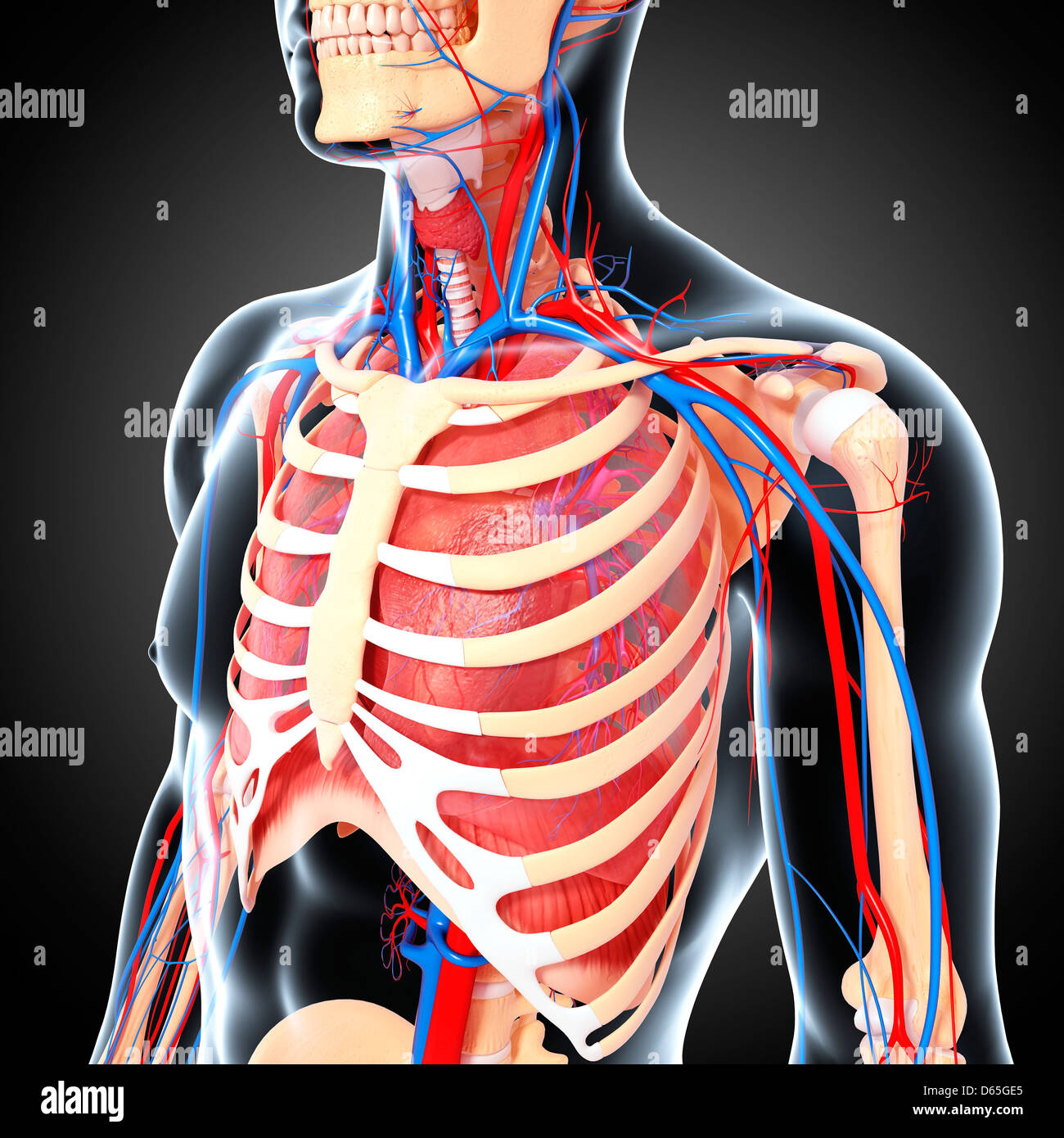
Upper Chest High Resolution Stock Photography And Images Alamy from c8.alamy.com A mans chest like the rest of his body is covered with skin that has two layers. Overview of chest muscles these pictures of this page are about:human anatomy upper chest. Nerves of the chest and upper back. 8 best upper chest exercises. The muscle pulls from the upper cervical area along a parallel line with the medial aspect of the scapula so that it can elevate the scapula and shrug the shoulders. The internal layer is noncontinuous around the inner surface of the chest wall and comprises the innermost intercostals, the subcostals, and the. Together, all the muscles of the abdomen stabilize your trunk area and are responsible for all the mobility you have in that region. • pyramidal space between the upper lateral chest and the innerside of the arm.
Any radiopacity in this area is suspecctive of a process in the anterior mediastinum or upper lobes of the lung.
Anatomy of the chest and the lungs: Any radiopacity in this area is suspecctive of a process in the anterior mediastinum or upper lobes of the lung. Anatomy of peritoneum and mesentery. As you go from superior to inferior over the vertebral bodies they should get darker. Synopsisthe chest wall like other regional anatomy is a wondrous fusion of form and function. • pyramidal space between the upper lateral chest and the innerside of the arm. Thanks for reading my anatomical guide to training! Anatomy of the chest area. Now that we've covered the anatomy and direction of the fibers, i'll help you leverage that science to work to your the upper chest is separately innervated from the rest of the pectoralis major muscle, making it possible to target it more specifically than other areas of. It is where the left ventricle hits against the chest wall. Heart labeled within womans chest stock. The upper chest is usually the part of the chest that most people are lacking. Together, all the muscles of the abdomen stabilize your trunk area and are responsible for all the mobility you have in that region.
Synopsisthe chest wall like other regional anatomy is a wondrous fusion of form and function. The upper posterior border of the heart is formed by the left atrium. Anatomy is to physiology as geography is to history: Understanding chest wall anatomy is paramount to any surgical procedure regarding the chest and is vital to any reco. Upper division of left superior lobar bronchus.
Abdomen Wikipedia from upload.wikimedia.org Apical, posterior and place one hand on top of the other affected over area or place one hand place one and on each side. Located at the level of the intervertebral disc between t4 and t5. Branches of the radial and ulnar arteries muscles: The upper chest is usually the part of the chest that most people are lacking. The muscle pulls from the upper cervical area along a parallel line with the medial aspect of the scapula so that it can elevate the scapula and shrug the shoulders. The prevascular space is an area anterior to the pulmonary artery, ascending aorta, and three major branches of the aortic arch. It is where the left ventricle hits against the chest wall. Anatomy of the chest & abdomen.
Heart labeled within womans chest stock.
It is where the left ventricle hits against the chest wall. 8 best upper chest exercises. Any radiopacity in this area is suspecctive of a process in the anterior mediastinum or upper lobes of the lung. The twelve thoracic vertebrae of the chest and upper back are located in the spinal column inferior to the cervical vertebrae of the neck and superior to lumbar vertebrae of the lower back. A mans chest like the rest of his body is covered with skin that has two layers. Anatomy is to physiology as geography is to history: Master upper extremity anatomy by learning about all its bones, muscles, arteries, and nerves at kenhub. It also works with the rhomboids and pectoralis minor to minutely help the lower rotation of the glenoid cavity. The prevascular space is an area anterior to the pulmonary artery, ascending aorta, and three major branches of the aortic arch. Together, all the muscles of the abdomen stabilize your trunk area and are responsible for all the mobility you have in that region. Upper back pain and chest pain can occur together. Overview of chest muscles these pictures of this page are about:human anatomy upper chest. Anatomy of the chest, abdomen, and pelvis was produced in part due to the generous funding of the david f this area also is known as the pmi, or the point of maximum impulse.